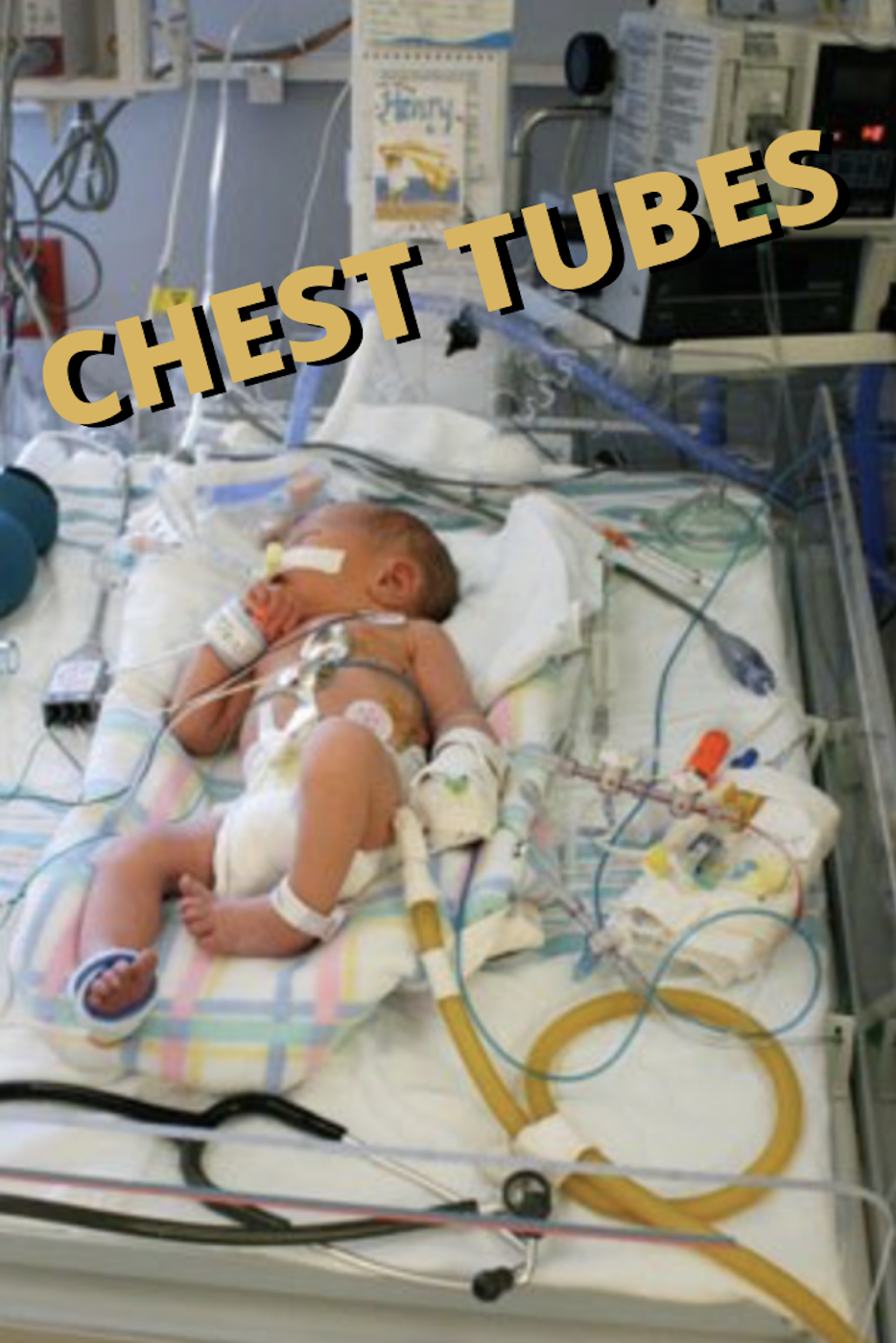
There were many subjects that I couldn’t quite wrap my brain around during nursing school. One of them being CHEST TUBES! I remember feeling so dumb and embarrassed in class. Everyone else understood the concept just fine whereas I couldn’t comprehend it for the life of me. “Is bubbling good or bad?” “Is the water seal chamber on the right or left?” So. Much. PTSD. Good times… It wasn’t until I actually started working with them on a daily basis in the NICU where it all finally clicked and the lightbulb turned on.
WHY ARE CHEST TUBES PLACED?
Chest tubes are placed in babies as a means to provide relief of respiratory distress associated with various conditions & etiologies.
Clinical indications for a chest tube include:
Pneumothorax. An air leak in the pleural space, causing the lung to collapse due to compression.
Hemothorax. The same as above but with blood in the pleural space instead of air.
Empyema. A collection of pus in the pleural cavity.
Pleural effusion. Fluid excess in the pleural space.
Chylothorax. An accumulation of chyle in the pleural space.
Post operatively as a surgical drain following cardiac surgery, a thoracotomy, or mediastinal surgery.
How does a chest tube work?
A chest tube is placed in the pleural or mediastinal space in order to restore the negative intrathoracic pressure needed for lung re-inflation. This involves draining/removing any fluid, blood or air that has accumulated and may be compressing the lung(s). The tube is connected to a drainage system that uses gravity and/or suction to remove the foreign substance and to prevent the air from re-entering. This helps with re-expansion of the lung(s) and helps to restore the negative pressure in the pleural space. Without intervention, this may lead to serious respiratory or circulatory compromise and rapid deterioration of the baby.
WHAT ARE THE COMPONENTS OF A CHEST TUBE?
Drainage collection chamber. This monitors the drainage color and amount. Depending on the age and size of your baby, the normal hourly output varies. This chamber is on the right side, just below the tube that is coming from the patient.
Water seal chamber. Water will fluctuate with inspiration and expiration. This chamber must be filled and maintained at the 2cm level to ensure proper operation. It should not be overfilled, as it is more difficult to get air out of the pleural space when this occurs. The water-seal chamber serves as a one-way mechanical valve that allows air to leave the chest and prevents it from re-entering the patient. A small amount of intermittent bubbling is normal. Continuous, turbulent bubbling can indicate an air leak.
—> Note: If water does not fluctuate, the lung has either re-expanded or there is a kink.
Air leak monitor. Excessive bubbling in the tubing proximal to the baby (i.e. the tubing coming out from the insertion site) indicates an air leak. It could be that the occlusive dressing has lifted and air is entering through the chest incision. If your patient has a pneumothorax, intermittent bubbling may be seen and is considered a normal finding.
Suction control chamber. This is filled with water. Remember, the height of water is what controls the amount of suction! Bubbling is expected in this chamber as it indicates that an appropriate amount of suction is being used. This chamber is located on the left, just below the tube that is connected to the wall suction.
TROUBLESHOOTING TIPS
There are many types of chest tubes, including percutaneously inserted pigtails, or larger, surgically placed chest tubes. Regardless of the type or brand, great care and caution should be taken as they are very flexible and prone to kinking, clotting, and/or dislodging!
It is important to avoid kinks or pressure in the tubing. In nursing school, they drill in your brain the importance of not milking or stripping the tubing. This is a true statement! Unless you have an MD order, only the physician or surgeon can do this. Also, do not lift the drainage system above the patient’s chest because the fluid may backflow into the pleural space. If the tube becomes dislodged, cover the area with a sterile dressing taped 3 sides down so that air can escape but not enter.
In order to prevent accidental disconnection and/or contamination, secure the tubing to the bed linens (with a rubber band & safety pin) to ensure it has a straight flow to the collection chamber and has no dependent loops or kinks. The chamber should be taped to the floor in an upright position. NEVER clamp a chest tube and do not pinch/occlude it when checking patency!
COMPLICATIONS
There are many complications to watch for following chest tube placement. Some of these include:
A tension pneumothorax. May be caused by a clamped chest tube. Signs & symptoms include mediastinal shift to the unaffected side, reduced venous return, increased respiratory distress, dysrhythmias, diaphoresis, tachycardia, hypotension, absent breath sounds on the affected side, chest pain, and dry cough. Tracheal deviation is a late sign and may not always be present.
Hypovolemic shock from excessive chest tube drainage. This can occur if the fluid replacement is not enough to meet the baby’s needs. Signs & symptoms include hypotension, tachycardia, diaphoresis, cool skin, and decreased capillary refill.
Air leak. Signs & symptoms of an air leak within the closed system include absence of drainage, absence of fluctuations in the water-seal chamber, or continuous bubbling in the water-seal chamber.
What is Tidaling?
Fluctuations in the fluid level (tidaling) occur when pressure changes in the pleural space (e.g. when the patient breathes). During inspiration, as negative pressure increases, so does the water level. During expiration, negative pressure in the pleural space decreases as does the water level. If a patient is on a mechanical ventilator, you will observe the opposite effect in the water column. This is due to the positive pressure inside the lungs applied by the vent instead of the negative pressure that normally occurs with non-assisted breathing.
Chest Tube Management
After the MD places the order, the first thing you should do is gather all of the supplies and prepare your collection container.
Fill the water-seal chamber with the prefilled syringe that comes in the package of the chest tube drainage system. This will allow the detection of air leaks. However, if the chamber is not filled, the system is still sealed.
Maintain the chamber in an up-right, secure position. It may be best to tape it to the ground or hang it on your patient’s bed frame… NOT THE RAILING!
For continuous suction, set the dial in the drainage system to the cm H2O ordered by the MD.
Set the wall suction to 80-120 mmHg. When suction is applied to a closed chest drainage system and attached to the patient, the orange bellow indicator on the front of the collection chamber will expand past the indicator mark on the chamber.
The regulator on the system is pre-set to -20 cm H20. This can be adjusted via turning the rotary dial to the setting ordered.
For water seal, no suction is necessary. To place a chest tube to water seal from suction, simply turn off the wall suction. In other words, the system is considered to be at water seal if it is not attached to suction or if the suction is turned off. You DO NOT need to disconnect the drainage system from the wall suction. If your patient does not tolerate water seal and needs to be restarted on suction, your tubing is already connected and ready to go. Some patients may need to be restarted on suction if the chest x-ray demonstrates recurrence of the problem.
The initial occlusive dressing should be placed by the MD. However, if the dressing becomes loose, saturated, soiled or begins lifting, an RN is able to perform the subsequent dressing changes as long as there’s an order. The occlusive dressing consists of two split drain sponges around the chest tube, followed by a gauze dress on top and then a semipermeable transparent dressing over that (example: tegaderm). This ensures a closed, sterile drainage system!

